Common Ankle & Foot Disorders Comprehensive Diagnosis & Treatment
Introduction A solid understanding of anatomy is essential to effectively diagnose and treat patients with foot and ankle problems. Anatomy is a road map. Most structures in the foot are fairly superficial and can be easily palpated. Anatomical structures (tendons, bones, joints, etc) tend to hurt exactly where they are injured or inflamed.

Ankle Anatomy Sport Med School
The Basics of Ankle Anatomy and Foot Anatomy Basic anatomy for any joint structure within the body includes bones, joints, muscles, tendons, and ligaments. For our purposes, we will be discussing Ankle Anatomy and Foot Anatomy structures specifically. Terms to Know: Lateral: outside Posterior: backside Anterior: frontside Medial: inside
Anatomy of the Foot and Ankle Astoria Foot and Ankle Surgery
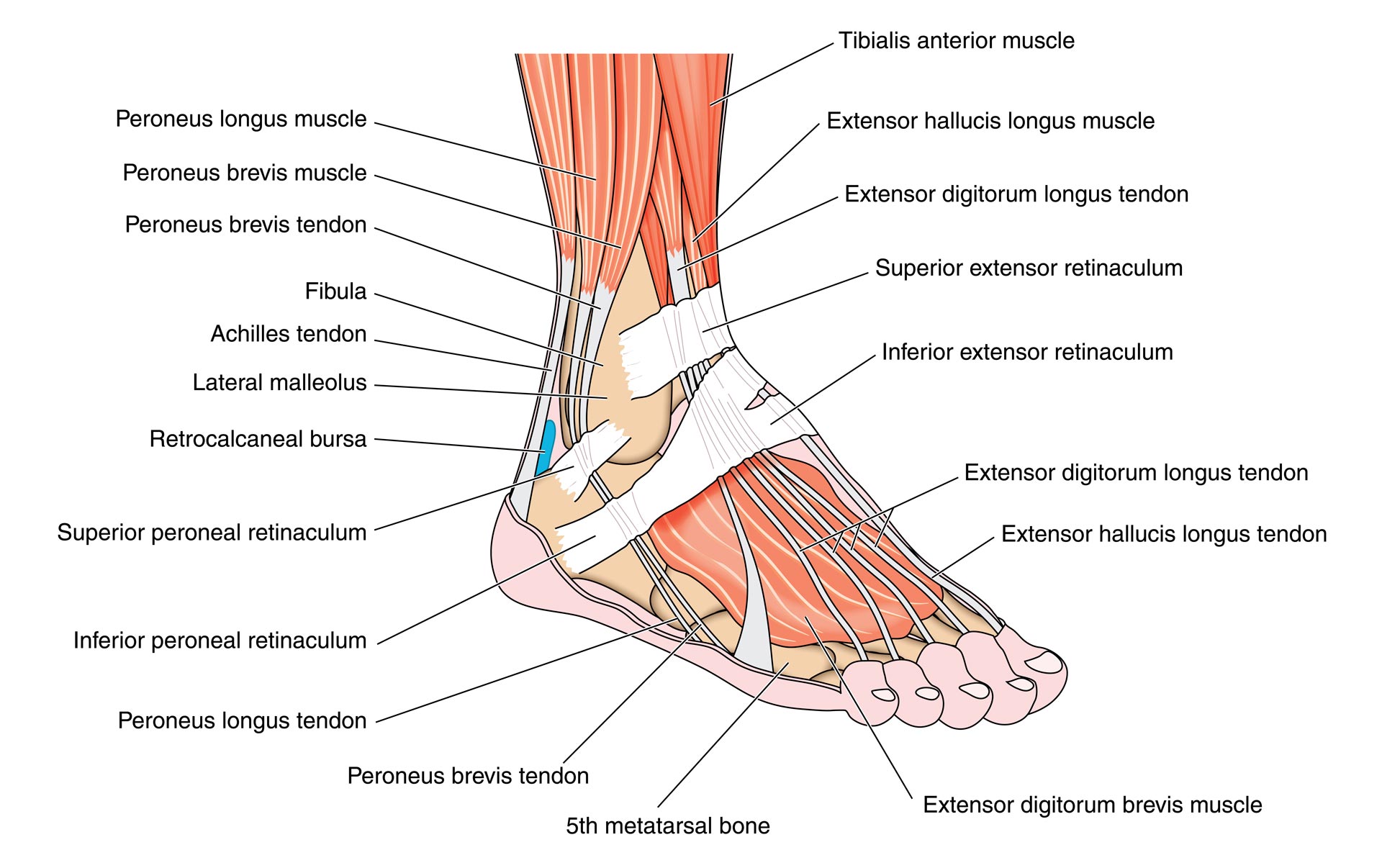
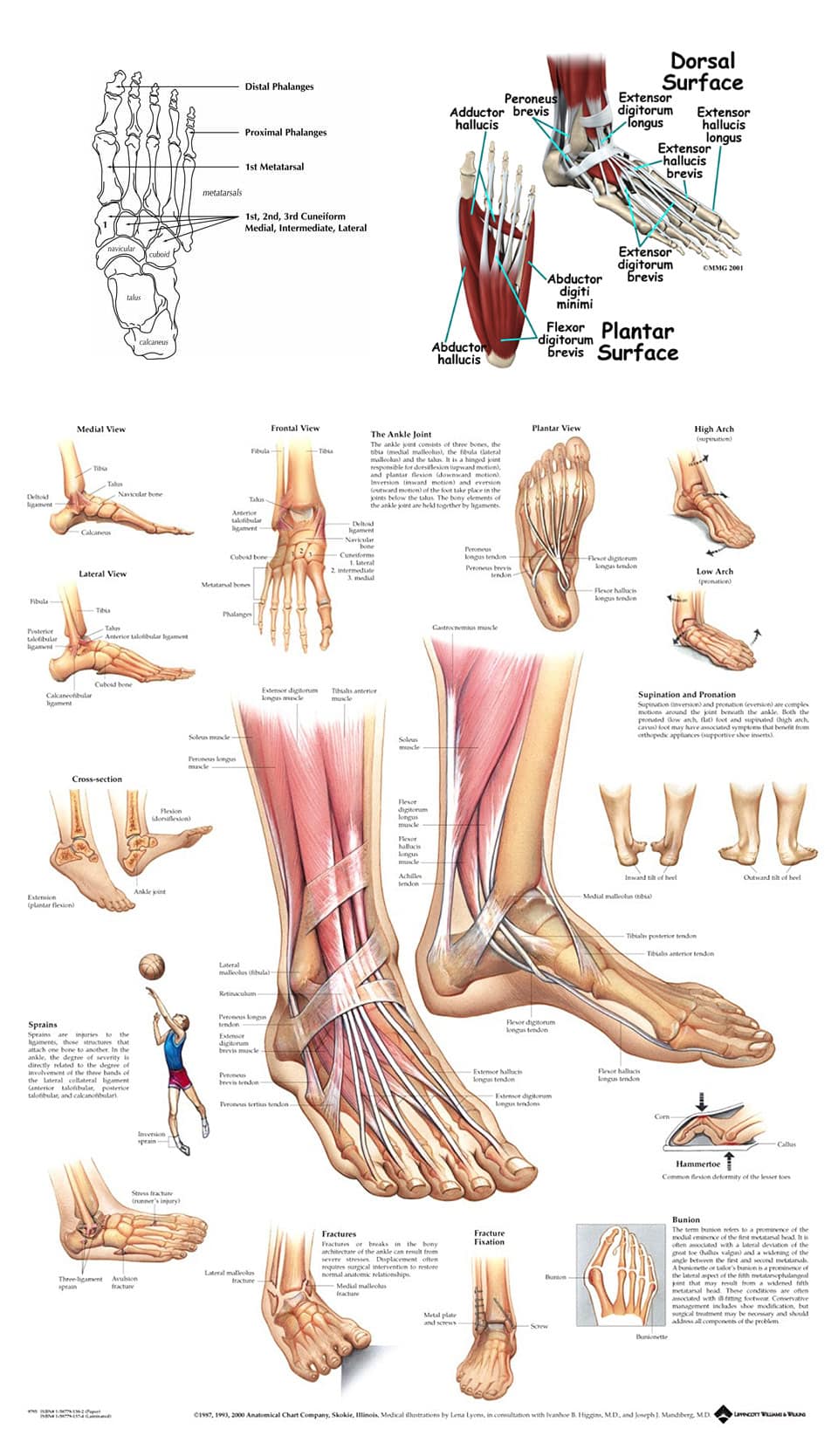
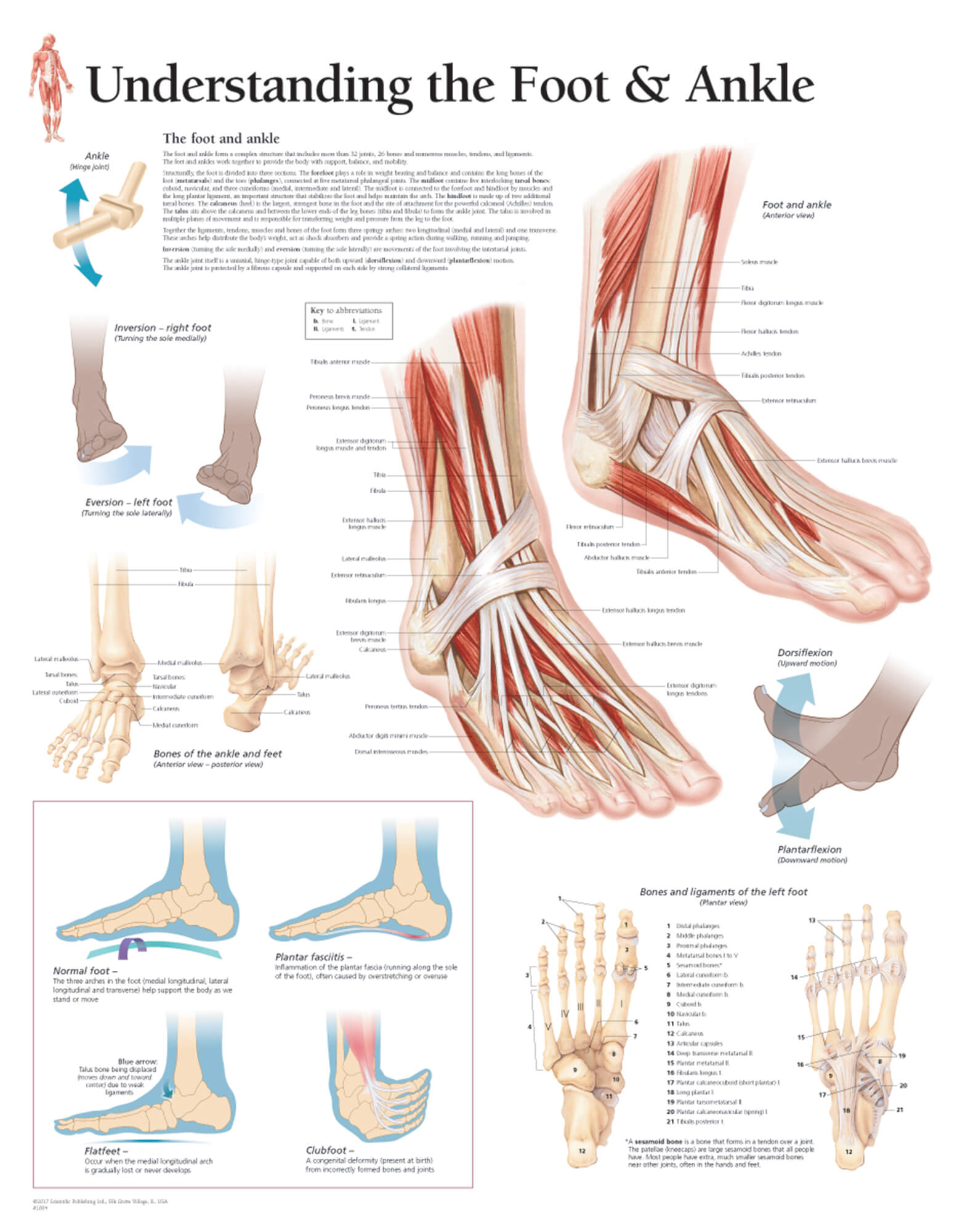
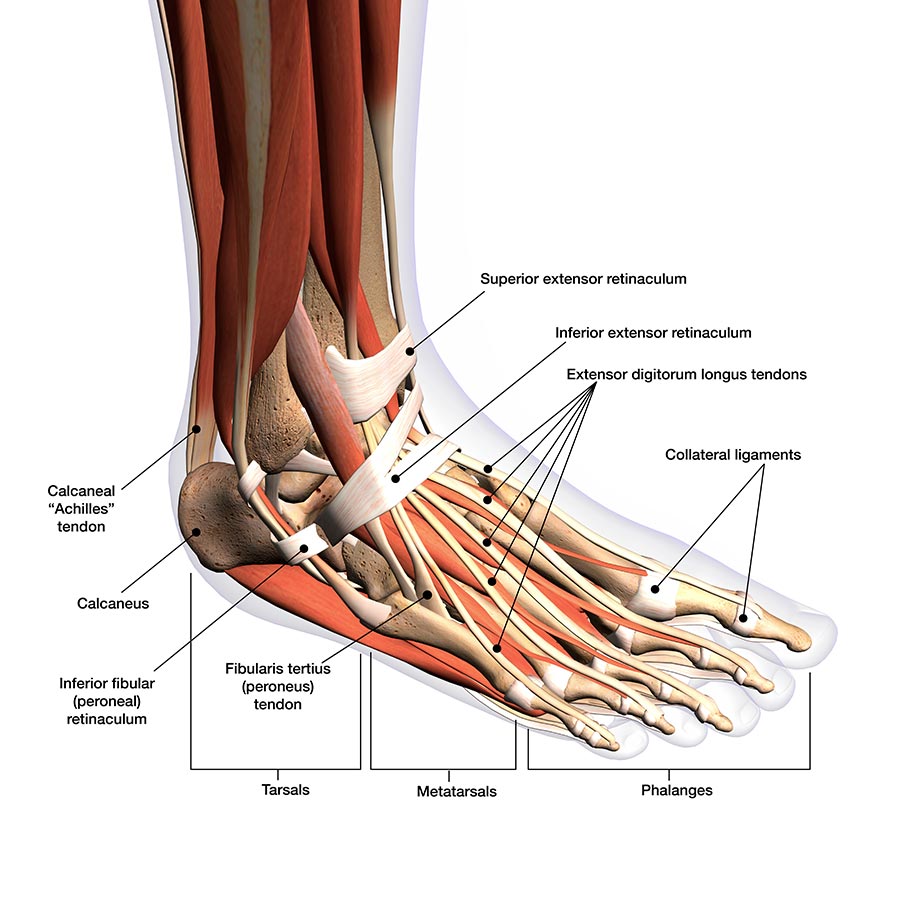
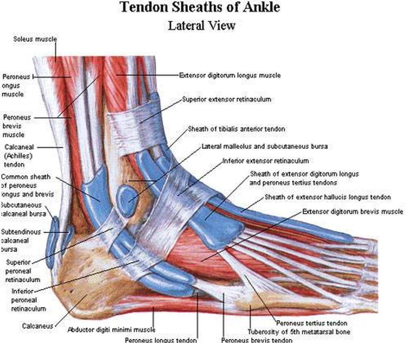
The foot and ankle form a complex system which consists of 28 bones, 33 joints, 112 ligaments, controlled by 13 extrinsic and 21 intrinsic muscles. The foot is subdivided into the rearfoot, midfoot, and forefoot. It functions as a rigid structure for weight bearing and it can also function as a flexible structure to conform to uneven terrain.

Anatomy of the Ankle Elliot's Blog
It is made up of over 100 moving parts - bones, muscles, tendons, and ligaments designed to allow the foot to balance the body's weight on just two legs and support such diverse actions as running, jumping, climbing, and walking. Because they are so complicated, human feet can be especially prone to injury.

This chart shows foot and ankle bone and ligament anatomy, normal movement of the joints, and
Function What does the ankle joint do? Your ankles bend and flex anytime you're moving to keep you stable and maintain your balance. Your ankles move in two directions: Plantar flexion: Down, away from your body. Dorsiflexion: Up, toward your body. Anatomy Where is the ankle joint located? The ankle is at the lower end of your leg.
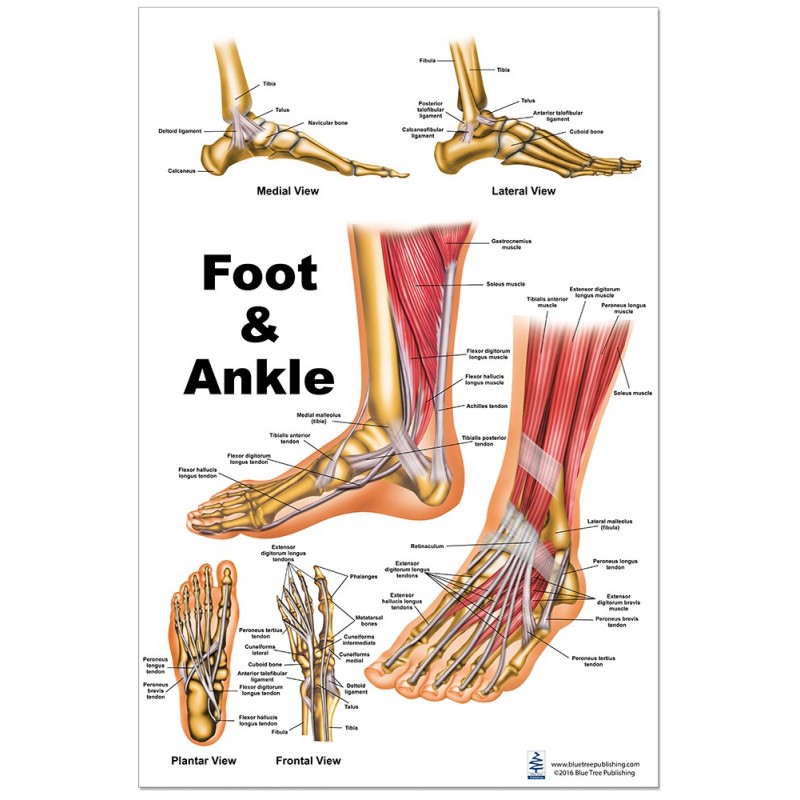
diagram of foot and ankle
Foot and ankle anatomy consists of 33 bones, 26 joints and over a hundred muscles, ligaments and tendons. This complex network of structures fit and work together to bear weight, allow movement and provide a stable base for us to stand and move on.
Ankle impingement syndrome causes, symptoms, diagnosis & treatment
The ankle joint or tibiotalar joint is formed where the top of the talus (the uppermost bone in the foot) and the tibia (shin bone) and fibula meet. The ankle joint is both a synovial joint and a hinge joint. Hinge joints typically allow for only one direction of motion much like a door-hinge.
Understanding the Foot & Ankle Scientific Publishing
In this animated episode of eOrthopodTV, orthopaedic surgeon Randale Sechrest, MD discusses the anatomy of the ankle joint.

Ankle and Foot Pain Massage Therapy Connections
The ankle joint, also known as the talocrural joint, allows dorsiflexion and plantar flexion of the foot. It is made up of three joints: upper ankle joint (tibiotarsal), talocalcaneonavicular, and subtalar joints. The last two together are called the lower ankle joint.

Foot and Ankle Musculoskeletal Key
The talus, or ankle bone: The talus is the bone at the top of the foot. It connects with the tibia and fibula bones of the lower leg. The calcaneus, or heel bone: The calcaneus is largest of.

ankle anatomy Health ankle anatomyankle anatomy
3.9K Share 747K views 11 years ago Foot/ Anatomy Dr. Ebraheim's educational animated video describes anatomical structures of the foot and ankle, The Bony Anatomy, The Joints, Ligaments,.

Muscles that lift the Arches of the Feet
The ankle and foot are anatomically complex areas with a broad spectrum of intra- and extra-articular pathologies. This chapter reviews basic anatomical features and gives an overview on common pathologic conditions with an emphasis on trauma/sports injuries and MR imaging. Keywords:
Foot and ankle anatomy, conditions and treatments
25K Share 2.2M views 11 years ago Animated Orthopedic Anatomy Tutorials In this episode of eOrthopodTV, orthopaedic surgeon Randale C. Sechrest, MD narrates an animated tutorial of the anatomy.
Pictures Of Ankle MusclesHealthiack
Anatomy of the foot and ankle. Arthritis Foundation. Anatomy of the foot. PHED 301 Students BC Campus. Advanced Anatomy 2nd. Ed.: The Foot. Bito T, Tashiro Y, Suzuki Y, et al. Forefoot transverse arch height asymmetry is associated with foot injuries in athletes participating in college track events.
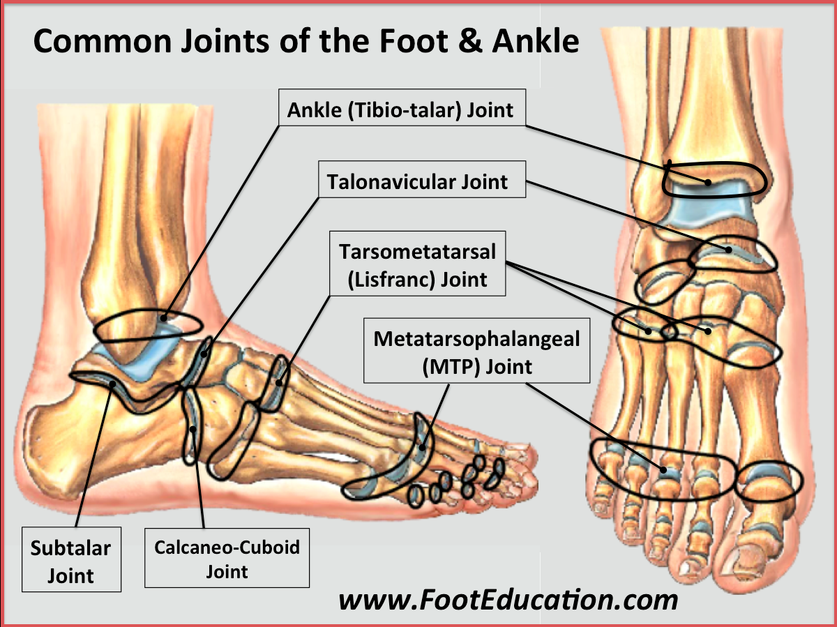
Bones and Joints of the Foot and Ankle Overview FootEducation
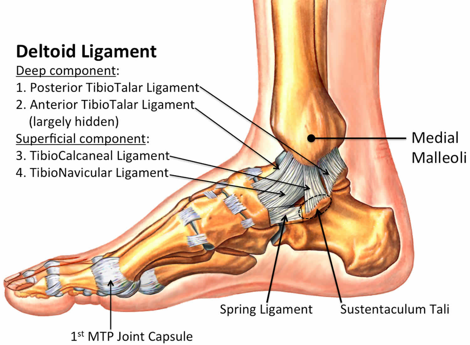
The ankle joint is formed by three bones; the tibia and fibula of the leg, and the talus of the foot: The tibia and fibula are bound together by strong tibiofibular ligaments. Together, they form a bracket shaped socket, covered in hyaline cartilage. This socket is known as a mortise.
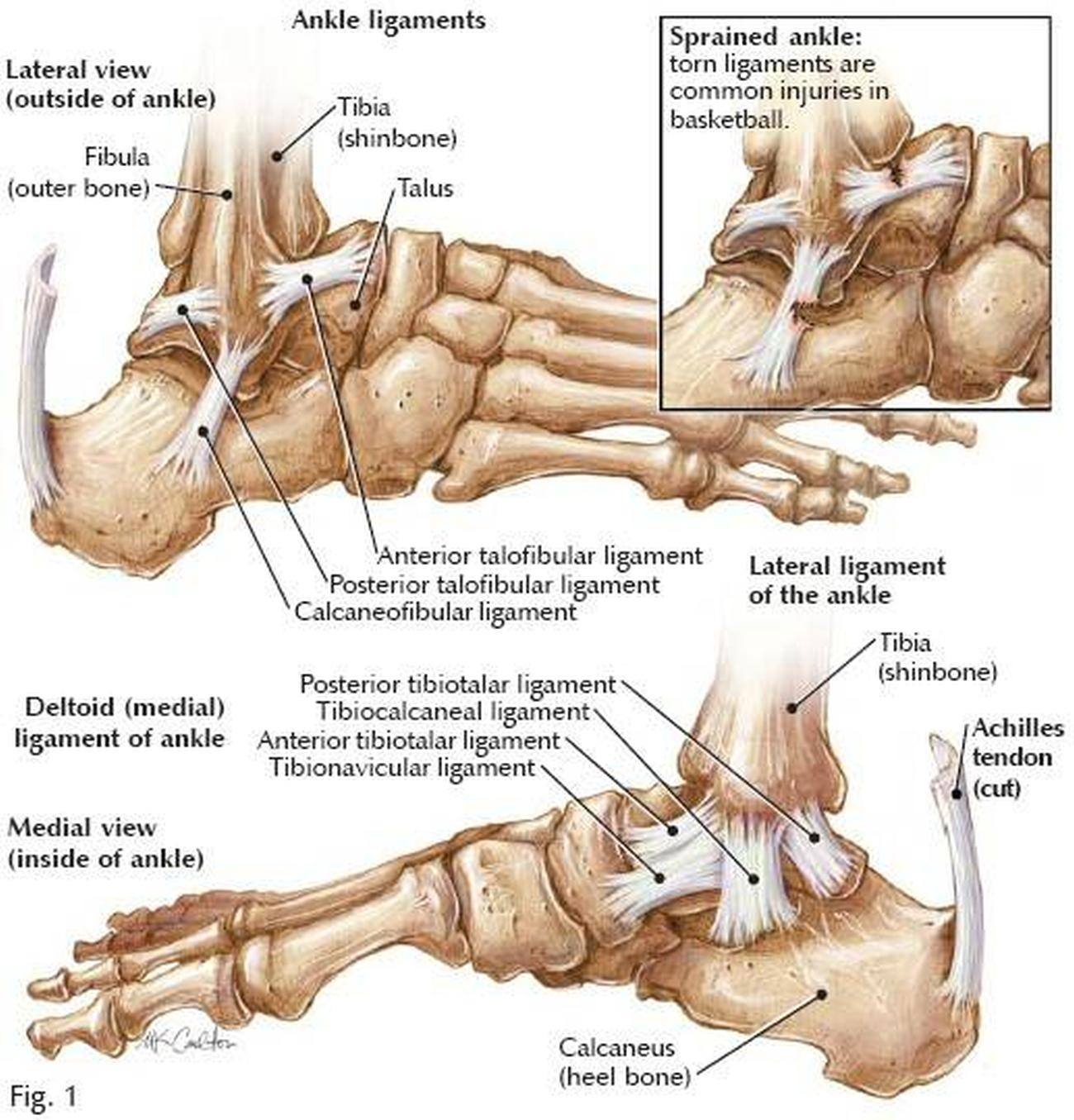
Pictures Of Ankle Joint Ligaments
Ligaments of the Foot and Ankle. Tell us where the pain is. Use our interactive tool. Use our Anatomy tools to learn about bones, joints, ligaments, and muscles of the foot and ankle. FootEducation is committed to helping educate patients about foot and ankle conditions by providing high quality, accurate, and easy to understand information.
